Wildlife Rescue And Rehabilitation Wound Care
An injured hawk surviving a brutal vehicle strike often dies hours later inside a quiet, sterile clinic room. The physical trauma spares the bird, but the sheer physiological terror of human handling causes a fatal biological collapse. Effective wildlife rescue and rehabilitation hinges entirely on controlling these physiological variables during the first vital hours of care. Rescuers face an unforgiving environment where standard veterinary practices routinely fail wild patients. Proper animal first aid serves as the mandatory bridge between a violent field injury and long-term clinical recovery, stabilizing fragile systems before irreversible shock sets in. Clinical professionals must execute flawlessly, managing raging inflammation cascades while preventing deadly stress responses. Perfecting these advanced wound care techniques turns basic compassionate interventions into highly scientific procedures, ensuring injured wild patients survive their trauma, heal completely, and safely return.
The Golden Rules of wildlife rescue and rehabilitation wound care
Treating a wild patient completely defies the logic governing domestic veterinary care. Domestic pets seek comfort from human touch, whereas wild prey and predators view every clinical intervention as an imminent lethal threat. This situation forces specialists to adopt strict golden rules within wildlife rescue and rehabilitation. Practitioners immediately classify all trauma-related wounds as heavily contaminated upon presentation, demanding instant, aggressive irrigation protocols.
According to guidelines published by the Washington Department of Fish and Wildlife, handlers must handle and interact with wildlife in rehabilitation facilities as little as possible, restricting contact strictly to providing quality husbandry and necessary medical treatment. A domestic dog will tolerate a thirty-minute examination, but a wild fox will experience cascading organ failure under the exact same duration of restraint. Therefore, wildlife professionals operate with ruthless speed. They secure the patient, assess life-threatening deficits, and begin immediate stabilization before the animal comprehends the capture. Achieving full functional recovery requires flawless execution of these vital non-negotiable foundational handling principles every single time.
Prioritizing rescuer and patient safety
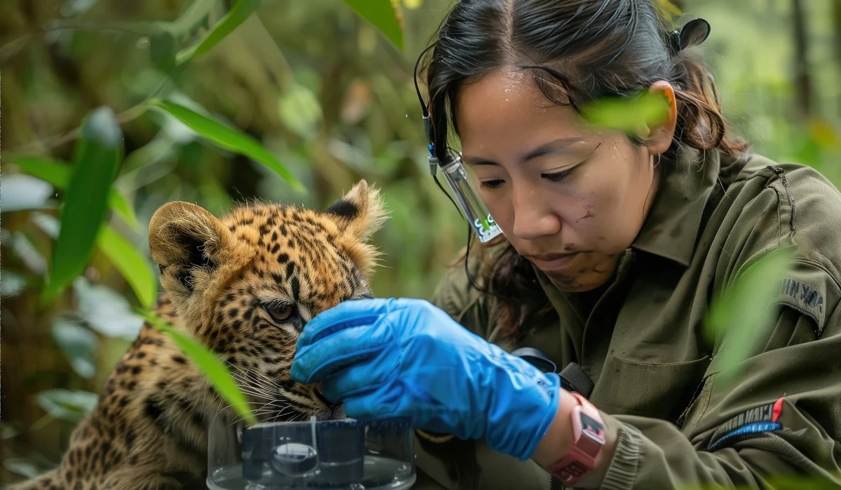
Approaching an injured wild animal demands highly specialized situational awareness and correct personal protective equipment. First responders wear heavy leather gauntlets and use large towels to establish physical barriers between themselves and snapping jaws or slicing talons. A cornered hawk or frantic bobcat perceives a towering human as an apex predator launching an attack. To neutralize this perception, the rescuer actively lowers their body posture to minimize their vertical silhouette. Furthermore, handlers strictly avoid direct eye contact, since wild animals universally interpret a sustained stare as a precursor to a lethal strike. Responders control their physical approach and utilize sturdy physical barriers to protect their own hands and faces from devastating lacerations. Simultaneously, this calculated approach protects the fragile patient from inflicting further skeletal damage upon itself while thrashing blindly against a poorly executed restraint attempt in the chaotic field environment.
Classifying wound severity in the field
Once responders secure the animal, they must rapidly classify the localized tissue damage to establish correct triage priorities. Rehabilitators quickly differentiate superficial abrasions from deep punctures, massive lacerations, or catastrophic degloving injuries. Degloving injuries occur when mechanical force entirely strips the skin away from the exposed muscle, leaving bone and vital tissues completely exposed to environmental pathogens. Meanwhile, seemingly minor puncture wounds from predator teeth frequently inject deadly bacterial loads deep into the fascia, requiring far more aggressive systemic treatment than a broad surface scrape. Wildlife medical teams rely heavily on the A.C.R.A.S.H. P.L.A.N. mnemonic—evaluating Airway, Cardiovascular, Respiratory, Abdomen, Spine, Head, Pelvis, Limbs, Arteries, and Nerves. This rigid framework ensures clinicians address neurological and cardiovascular deficits first, preventing them from developing tunnel vision on a gruesome but non-lethal laceration while the animal slowly asphyxiates.
Initial Assessment and Essential animal first aid
Immediate intake procedures dictate whether a critically injured wild patient lives or dies within the first hour. Before professionals even consider flushing a contaminated wound, they must execute rigorous animal first aid to combat systemic collapse. Physical trauma universally compromises vascular volume and initiates severe hemorrhagic shock. A rehabilitator rapidly checks the patient for active arterial bleeding, catastrophic bone fractures, and drastically altered mentation. During this intake phase, identifying and mitigating shock takes absolute precedence over localized wound management. Wildlife shock manifests rapidly, featuring pale mucous membranes, dilated pupils, and dangerously weak pulses that often beat two to three times per second in smaller species. Clinicians completely disregard the superficial injuries until they restore adequate blood pressure, administer vital fluids, and secure the airway, effectively buying the compromised animal enough time to survive further medical examinations.
The triage timeline
Executing a disciplined triage timeline maximizes the survival odds for traumatized wildlife. Handlers strictly limit the initial hands-on examination to a brutally short two-to-five-minute window. Within the first five minutes, professionals assess the airway, control arterial hemorrhage, and place the animal inside a dark, heated incubator to mitigate environmental stress. Over the next fifteen minutes, clinical teams prepare necessary medications, calculate exact fluid requirements based on precise body weight, and gather specialized wound care supplies outside the patient’s sight. At the sixty-minute mark, responders administer warmed subcutaneous fluids and powerful analgesics to stabilize the crashing metabolic system. Prolonging the initial visual examination beyond five minutes invariably spikes the patient’s heart rate, exacerbating shock and pushing a savable animal past the point of physiological recovery. Strict adherence to this rapid timeline consistently prevents catastrophic stress-induced fatalities.
Stabilizing the patient for transport
Transporting a compromised patient correctly requires careful environmental control and thermal support. Captured animals suffering from traumatic hyperthermia, where body temperatures easily exceed forty-one degrees Celsius, urgently need portable mist sprayers and an immediate intravenous infusion of cold Ringer’s lactate. Conversely, hypothermic animals require external warming discs placed securely underneath thick towels to gradually elevate their core temperature. Bystanders observing these critical interventions frequently ask what people should do when finding an injured wild animal.
According to guidelines published by Ventura County Animal Services, finders must place the animal in a dark, warm, quiet, and private transport space; therefore, the best approach involves covering the animal with a towel to minimize visual stress, placing it inside a well-ventilated carrier, and immediately transporting it to a licensed professional. This specific transport protocol definitively reduces the risk of deadly shock and ensures the patient survives the stressful transport long enough to receive dedicated, life-saving wound care at the clinic.
Flushing and Cleaning the Core of wildlife rescue and rehabilitation
Once the animal stabilizes, practitioners shift their focus to the physical process of proper wound irrigation, which forms the medical core of wildlife rescue and rehabilitation. Generating the exact correct physical pressure determines whether the tissue heals or dies. According to clinical guidelines published in the National Center for Biotechnology Information, the medical standard for safe, low-pressure wound irrigation ranges between four and fifteen pounds per square inch.
To perfectly generate this ideal pressure, rehabilitators utilize a 35-milliliter piston syringe equipped with an eighteen-gauge needle. This specific configuration delivers exactly seven to eight pounds of pressure per square inch. The same guidelines warn that irrigation pressures exceeding fifteen pounds per square inch cause wound trauma and forcefully drive surface bacteria deeper into exposed bone and fragile soft tissue, guaranteeing severe secondary infections. Simple free-drip flushing methods generate less than four pounds of pressure and completely fail to dislodge embedded microscopic debris. Perfecting this mechanical flushing technique drastically reduces healing times.
Selecting safe irrigation solutions
Choosing the correct chemical solution prevents iatrogenic damage to the delicate exposed cellular structures. Veterinarians rely heavily on a highly diluted 0.05 percent chlorhexidine diacetate solution or standard 0.9 percent normal sterile saline to flush open lacerations safely. These specific fluids eliminate heavy pathogen loads without destroying the fragile fibroblasts necessary for new skin generation. In sharp contrast, applying a standard 4 percent chlorhexidine surgical scrub directly into an open cavity acts as a potent cellular toxin, instantly killing exposed healthy tissue and halting the natural healing progression.
Furthermore, trained rehabilitators strictly ban harsh household chemicals like hydrogen peroxide from the treatment room. Research published in PubMed confirms that hydrogen peroxide and iodine serve as poor choices because they retard the contribution of fibroblasts to the healing process, aggressively destroying healthy granulating cells alongside the bacteria, turning a manageable shallow laceration into a deeply necrotic hazard. Applying precise, scientifically backed irrigation solutions preserves every microscopic millimeter of viable tissue.
Gentle debridement techniques
Following aggressive irrigation, clinicians perform gentle debridement to remove remaining necrotic tissue and stubbornly embedded foreign debris. This highly precise step requires immense patience, as the practitioner must carefully separate dead organic matter from the healthy granulation tissue directly underneath. Skilled rehabilitators use sterile thumb forceps and specialized curved scissors to snip away devitalized black margins without initiating fresh capillary bleeding. Scraping the wound bed aggressively tears the microscopic blood vessels, setting the entire healing process back by several days. If the trauma presents heavily embedded gravel or stubborn dirt, professionals utilize specialized wet-to-dry bandaging techniques rather than risking sharp surgical errors. The wet gauze actively pulls the microscopic debris upward as it slowly dries, performing a flawless, natural debridement that completely spares the vital exposed muscle fibers and accelerates the closure of massive gaps.
Advanced Bandaging for Diverse Anatomies
Wrapping wild patients introduces unique anatomical challenges that completely separate wildlife medicine from traditional dog and cat treatments. Securing a massive laceration on an owl requires vastly different spatial considerations than wrapping the crushed leg of an adult red fox. Improper bandaging materials readily restrict vital respiratory movements or permanently destroy the delicate waterproofing qualities of flight feathers, effectively dooming the patient even if the localized laceration heals perfectly. Consequently, exact material selection remains a non-negotiable staple of quality wildlife rescue and rehabilitation. Clinical teams utilize specialized cohesive wraps that contour tightly to bizarre limb shapes without slipping during frantic physical struggles in the enclosure. These flexible bandaging techniques must maintain rigid skeletal support while actively absorbing heavy exudate from the infected cavity, guaranteeing the injury site remains optimally protected against external environmental contaminants throughout the process.
Avian wing wraps and stabilization
Avian anatomy dictates highly specialized approaches to wing trauma. Research published by the National Center for Biotechnology Information highlights that unlike mammals, birds lack a thoracic diaphragm and a phrenic nerve, relying entirely on the mechanical expansion of their chest cavity to breathe. Therefore, wrapping a bandage entirely around a bird's torso will instantly cause fatal asphyxiation. To solve this physiological hurdle, rehabilitators apply the precise figure-eight wrap. This method weaves the bandage smoothly around the humerus and carpal joints, folding the damaged wing snugly against the body while leaving the sensitive chest completely uncompressed.
Practitioners successfully immobilize severe antebrachial fractures in raptors and delicate songbirds using this exact weaving pattern. A properly executed figure-eight bandage anchors the broken bones firmly in place, allowing the localized wound beneath to heal rapidly while granting the frightened bird full respiratory freedom to pant and vocalize naturally inside the intensive care unit during its extended clinical stay.
Mammalian limb and torso care
Mammals require radically different bandage structures to manage massive exudate production following a predator attack. Caretakers routinely apply wet-to-dry bandages over torn mammalian muscle tissue, utilizing sterile saline-soaked wide-mesh gauze to physically wick away the necrotic fluids. As beginners gather these specific materials, a common question arises regarding whether people can use human bandages on animals. While basic sterile gauze pads remain perfectly safe, responders must never apply adhesive human bandages to wild animals because the glue permanently rips out important fur and delicate feathers upon removal. Instead, wildlife professionals strictly rely on cohesive vet wrap that sticks exclusively to itself. This specialized material effectively secures the primary dressing to the limb without adhering to the surrounding coat, preventing devastating secondary skin avulsions when clinicians inevitably perform routine bandage changes later in the week to inspect the healing progress.
Managing Infection and Controlling Pain
Modern wildlife rescue and rehabilitation demands aggressive pharmacological intervention to conquer heavy bacterial loads and manage severe physical trauma. The antiquated clinical mentality suggesting wild creatures naturally tolerate excruciating pain better than domestic pets causes horrific suffering and spikes mortality rates. Today, specialists prioritize rapid pain mitigation and preemptive infection control as absolute pillars of the healing protocol. Treating a predator bite requires immediate systemic antibiotics because deadly Pasteurella bacteria rapidly multiply beneath the superficially healed skin surface.
Without these vital medications, localized wounds quickly cause catastrophic systemic sepsis. According to a study published in the National Center for Biotechnology Information, using sugar to treat wounds provides a safe, easy-to-teach, and cost-effective alternative modality; therefore, clinicians sometimes pack deep cavities with medical-grade granulated sugar to combat localized infections without heavily relying on antibiotics. The sugar naturally creates a powerful osmotic pull, forcefully drawing healing macrophages into the damaged area while rapidly desiccating and eliminating the deeply embedded bacteria lingering deep inside the tissue.
Recognizing signs of sepsis early
Identifying the clinical signs of systemic infection requires immense vigilance, especially since prey animals evolutionarily hide their symptoms to avoid attracting hungry predators. A rabbit suffering from overwhelming sepsis rarely displays overt whining or thrashing behaviors. Instead, handlers look for subtle but alarming indicators like severe lethargy, total anorexia, and drastically lowered body temperatures. An infected animal frequently sits hunched in the darkest corner of the enclosure, aggressively ignoring fresh food offerings while exhibiting labored, shallow breathing. Blood tests further confirm the bacterial battle, revealing skyrocketing white blood cell counts long before the localized wound displays gross purulent discharge. Intervening aggressively at the very first sign of these subtle behavioral changes determines whether the animal recovers smoothly or succumbs rapidly to an otherwise preventable massive secondary bacterial infection within forty-eight hours of initial medical intake.
Administering species-appropriate analgesics
Veterinarians rely on precise, species-appropriate analgesics to deliver safe, effective, multi-modal pain relief for severe trauma cases. Meloxicam, a highly effective nonsteroidal anti-inflammatory drug, drastically reduces painful swelling with minimal kidney side effects. For birds, practitioners typically dose Meloxicam between 0.2 and 0.5 milligrams per kilogram daily. Meanwhile, opiate efficacy varies wildly between different animal classifications. Clinicians heavily rely upon Butorphanol to provide consistent, powerful analgesia in injured avian patients. Conversely, Buprenorphine works flawlessly to eliminate severe pain in traumatized mammals, but it yields highly unpredictable and often completely ineffective results in avian species like hawks or parrots. Combining these carefully selected injectable NSAIDs and oral opiates creates a comprehensive pain management shield, empowering the patient to eat, rest, and heal comfortably despite carrying massively debilitating skeletal and muscular field injuries during their recovery.
Minimizing Capture Myopathy and Treatment Stress
Navigating the delicate balance between necessary medical treatment and stress reduction represents the true operational art of wildlife rescue and rehabilitation. A deer or heron can easily survive a horrific laceration but subsequently die from the sheer physiological terror of human handling. Veterinarians call this specific iatrogenic disease capture myopathy, which causes massive, irreversible muscle necrosis and devastating kidney failure driven by extreme physical exertion and unmitigated panic. Dark red myoglobinuric urine serves as the most glaring indicator of this fatal internal breakdown. Because no clinical cure exists for capture myopathy, preventative stress management becomes just as necessary as the physical bandage itself. Practitioners battle this phenomenon by strictly limiting auditory and visual stimuli, knowing that preventing terror ensures the resilient wild body maintains enough metabolic strength to actively heal the physical wounds effectively over time.
Creating a dark and quiet healing environment
Designing the perfect recovery enclosure requires eliminating all unnatural sensory inputs that cause instinctive prey responses. Handlers strategically place dense visual barriers around wire cages to block the animal from observing human movements across the room. Furthermore, eliminating artificial human noises completely turns the clinical setting into a tolerable healing space. Staff strictly forbid talking, playing radios, or loudly clanking metal instruments near the intensive care units. Even casual conversational tones register as highly aggressive predatory signaling to a stressed fox or injured hawk. Additionally, clinical teams systematically control the ambient temperature, utilizing environmental cooling to drastically multiply the animal's resistance to physical capture myopathy. Maintaining this dark, cool, absolutely quiet environment forces the patient's raging nervous system to finally decelerate, shifting valuable biological energy back toward aggressive cellular regeneration and wound closure.
Low-stress handling techniques
Executing physical treatments demands a highly choreographed approach to minimize terrifying restraint periods. Teams orchestrate every single medication draw, bandage preparation, and instrument layout outside the animal's visual range long before opening the cage door. Once fully prepared, handlers execute fast, firm hold techniques that secure the struggling patient instantly, completely eliminating frantic chasing around the enclosure. A skilled rehabilitator aims to complete the entire wound flushing, debridement, and re-bandaging process in under two minutes. Releasing the animal swiftly back into its dark enclosure abruptly terminates its escalating panic response. Prolonging this grip to admire the surgical work or perform unnecessary secondary checks exponentially increases the risk of sudden cardiac arrest. Absolute brevity during these physical encounters remains the most potent tool for keeping fragile, highly stressed wild creatures alive during ongoing rehabilitation.
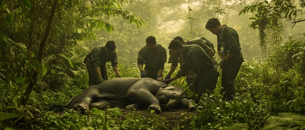
Connecting Individual Healing to species conservation
Careful clinical wound care naturally zooms out to generate a massive macro-level ecological effect. Healing a single endangered turtle, apex raptor, or mammal directly supports broader global species conservation initiatives. Returning healthy individuals back into small, isolated, or heavily inbred wild populations functions as a powerful tool known as genetic rescue. These rehabilitated animals outcross valuable new genetic diversity into fragile local groups, strengthening the entire biological lineage against future disease outbreaks or environmental shifts. Therefore, successfully patching a superficial laceration actively stabilizes declining habitats. When clinical professionals perfect their flushing pressures and administer exact pharmacological dosages, they effectively turn their solitary treatment tables into frontline battlegrounds for global preservation. Every perfectly executed wet-to-dry bandage ultimately helps secure the long-term biological viability of vulnerable populations worldwide against extinction.
The ripple effect of returning breeders to the wild
Releasing mature breeding adults back into their natural habitat creates an exponential biological ripple effect. Statistical population models clearly demonstrate that saving a large, sexually mature individual positively changes population survival far more than saving an infant. Florida sea turtle rehabilitation models, for instance, reveal that returning a single mature female to the ocean directly translates into thousands of future hatchlings over the ensuing decades. The grueling clinical labor invested in healing her propeller strike laceration directly secures the genetic lineage of her species. Every successful treatment ensures that apex predators continue regulating prey populations and that key herbivores continue dispersing essential seeds across vast forests. Saving one breeding animal effectively heals the entire connected environment, proving that high-quality medical intervention generates consequences extending far beyond the walls of the clinic to shape the future.
Tracking post-release survival rates
Empirical telemetry data rigorously proves that these intense clinical interventions actually translate to long-term wilderness survival. Biologists attach lightweight radio trackers and durable leg bands to recovering patients, monitoring their hunting success and movement patterns long after their release. A detailed study published in the National Center for Biotechnology Information tracking post-rehabilitation koalas demonstrated an impressive 58.5 percent long-term survival rate in the wild. When observing these extensive tracking efforts, many people ask whether wildlife rehabilitation actually helps conservation. Yes, rehabilitating and successfully releasing individuals helps maintain valuable genetic diversity and stabilizes fragile local populations. Gathering this hard data validates the grueling physical efforts required to carefully clean a contaminated wound or apply a flawless figure-eight bandage. Every single healed laceration and opened cage door contributes measurably and directly to the broader, ongoing survival of the targeted species in their natural habitat.
The Lifesaving Effect of wildlife rescue and rehabilitation
Treating a wild creature forces veterinary professionals to navigate a demanding biological minefield every single day. The entire process from executing flawless initial field triage to completing massive cellular wound debridement requires extraordinary technical precision. Practitioners successfully conquer severe physiological hurdles, actively managing deadly capture myopathy while fighting aggressive secondary bacterial infections with perfectly dosed medications. Integrating these advanced medical strategies ensures a highly elevated standard of care for patients that inherently hide their agonizing pain. Ultimately, dedicating resources to wildlife rescue and rehabilitation turns solitary clinical interventions into powerful global ecological victories. When rehabilitators successfully manage a severe fracture or clean a gruesome predator bite, they directly preserve the vital genetic diversity of fragile local environments. Perfecting these advanced wound care skills remains one of the most direct, hands-on methods for genuinely healing our natural world.
Recently Added
Categories
- Arts And Humanities
- Blog
- Business And Management
- Criminology
- Education
- Environment And Conservation
- Farming And Animal Care
- Geopolitics
- Lifestyle And Beauty
- Medicine And Science
- Mental Health
- Nutrition And Diet
- Religion And Spirituality
- Social Care And Health
- Sport And Fitness
- Technology
- Uncategorized
- Videos